What Is Foot Drop?
Foot drop is a general term used for difficulty lifting the foot upward at the ankle and bringing the toes up, from the ground toward the head. This type of injury results in a foot that essentially "hangs" at the ankle.
If you have foot drop, your foot might drag on the floor when you walk and tripping over the foot becomes a risk. You may have to raise your thigh high when you walk to help your foot clear the ground. You may also experience numbness on the skin of your leg and foot. Depending on the cause, foot drop can affect one or both feet. Foot drop isn't a disease, but it may be a sign of an underlying neurological or muscular problem. Sometimes foot drop is temporary, but it can be permanent. Fortunately, there are treatment options available.
What Causes Foot Drop?
Foot drop is caused by weakness or paralysis of the muscles involved in lifting the foot upward at the ankle and bringing the toes up, from the ground toward the head. A person who has foot drop may have difficulty walking and need to wear a brace on the leg called an ankle-foot orthosis or AFO. Possible causes of foot drop include lumbar disc herniation (damage to a nerve root in the lumbar spine), damage to the peroneal nerve (usually near the knee), damage to the nerve bundles in the lumbosacral plexus or to the sciatic nerve itself. A foot drop can begin after an injury to the back or leg, an operation on the hip or knee, or even such benign activities as squatting or crossing the legs for prolonged periods of time.
While the disorders described above refer to a “true” foot drop, conditions that cause spasticity which also result in difficulty lifting the foot are also sometimes referred to as foot drop. Disorders that affect the spinal cord or brain — such as multiple sclerosis, stroke, or brain injury— may cause foot drop. This foot drop is different from that described above, because it is primarily a result of spastic muscles pulling the foot down and in. Even if there is muscle function in the muscles that pull the foot and toes up, they cannot overcome these overactive muscles and a similar AFO brace is often prescribed.
(See spasticity for further information on this type of foot drop.)
Diagnosis of Foot Drop
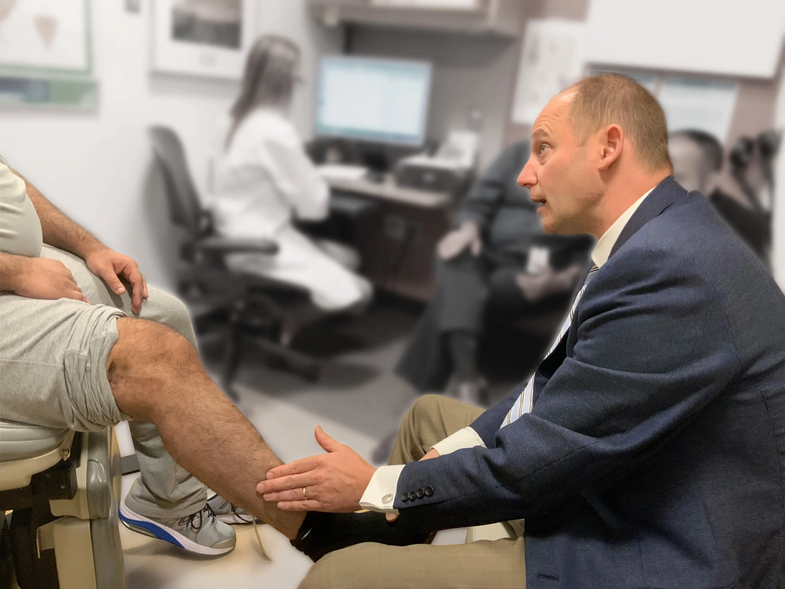
Foot drop is usually diagnosed during a physical exam. Your doctor will watch you walk and check your leg muscles for weakness. They will likely also check for numbness on your shin and on the top of your foot and toes. Foot drop is sometimes caused by bony narrowing in the spinal canal or by a tumor or cyst pressing on the nerve in the knee or spine. Imaging tests can help pinpoint these types of problems.
Electromyography (EMG) and nerve conduction studies measure electrical activity in the muscles and nerves. These tests can be uncomfortable, but they're useful in determining the location of the damage along the affected nerve. They can also help determine severity of damage and potential for recovery.
Treatment of Foot Drop
Treatment for foot drop depends on the cause. If the cause is successfully treated, foot drop might improve or disappear altogether. If the cause can't be treated or the foot drop persists in spite of that treatment, reconstruction may be required. Treatment for foot drop might include therapy, bracing or surgery.
Exercises that strengthen your leg muscles and help you maintain the range of motion in your knee and ankle might improve gait problems associated with foot drop. Stretching exercises are particularly important to prevent the stiffness in the heel or shortening of the achilles tendon. If this tendon shortens, pulling the foot up can become even more challenging. A brace on your ankle and foot or splint that fits into your shoe can help hold your foot in a normal position. Depending upon the cause, and if your foot drop occurred in recent months, nerve surgery might be helpful.
When the weakness is due to compression of the peroneal nerve, a simple operation can be performed to improve the situation. The peroneal nerve runs around the neck of bone on the outside of the leg (fibula) just below the knee. It then runs under a muscle that frequently has a tight fascial edge (the peroneus longus). At the point where the nerve runs under this muscle, this tight spot can be released and pressure eliminated. Many times, this is all that is required to restore function to the foot.
If weakness is due to nerve root compression within the lumbar spine, often an operation can be performed to open the space where the nerve leaves the spine (the spinal foramen) by either removing a herniated disk (microdiscectomy), opening this foramen (foraminotomy), or in more complex cases, a combination of these procedures with or without a fusion, where the bones are fixed together to avoid problematic movement.
Sometimes these procedures will not be enough to restore the function of the foot, which is when a nerve transfer can sometimes be used. This procedure involves taking "donor" nerves with less important roles — or branches of a nerve that perform redundant functions to other nerves — and "transferring" them to restore function in a more crucial nerve that has been severely damaged.
A nerve transfer can be performed in an attempt to correct a foot drop which may involve taking branches of the tibial nerve, the nerve that supplies muscles that push the foot down, and plugging those in (transferring them) to nerves that supply muscles involved in pulling the foot up. Either the branches of the tibial nerve that innervate the muscles that flex the toes or those that contribute to flexing the calf muscles and pushing the foot down may be used as donor nerves.
After this procedure, patients will still be able to push the foot down. However, as they regain function from the nerve transfer, they have to retrain their foot to use these muscles to pull the foot up. Recovery of function after nerve transfer is a process with initial results noticeable three to six months after surgery, with more full movement returning twelve months after surgery.
If foot drop is long-standing, your doctor might suggest a tendon transfer procedure that transfers a working muscle to a different part of the foot to provide the missing movement.
Watch video on Reversing Foot Drop in Athletes
Schedule a Consult with the Paralysis Center today (844) 930-1001.
Tips to help you get the most from a visit to the Paralysis Center
Before your visit, write down questions you want answered.
Bring someone with you to help you ask questions and remember what your Specialist tells you.
At the visit, write down the name of your diagnosis, and any new medicines, treatments, or tests. Also write down any new instructions your specialist gives you.
Know why a new medicine or treatment is prescribed, and how it will help you. Also know what the side effects are.
Ask if your condition can be treated in other ways.
Know why a test or procedure is recommended and what the results could mean.
Know what to expect if you do not take the medicine or have the test or procedure.
If you have a follow-up appointment, write down the date, time, and purpose for that visit.
Know how you can contact your Paralysis Specialist if you have questions.


